Veterinary Diagnosis of Salpingitis: A Complete Guide for Poultry Keepers

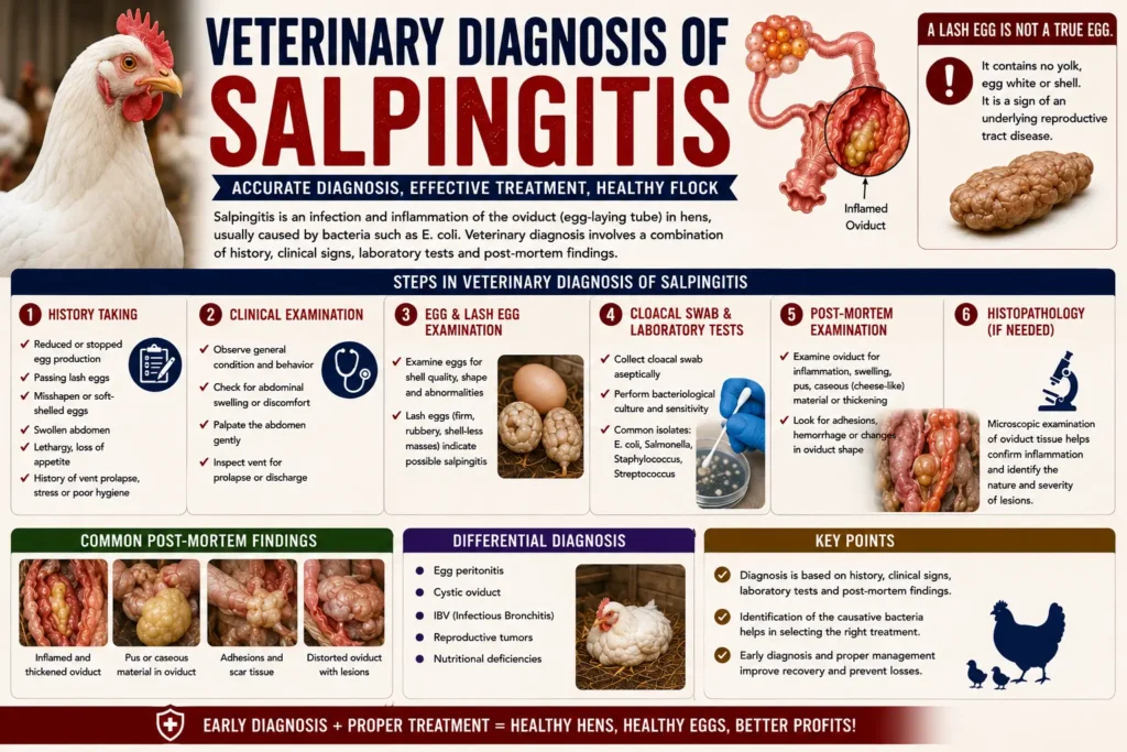
If your hen has stopped laying, sits hunched with a swollen belly, or walks like a penguin, salpingitis could be the reason. The veterinary diagnosis of salpingitis relies on a mix of physical exams, imaging, lab work, and sometimes necropsy, because the symptoms overlap with several other reproductive diseases. This guide walks through exactly how vets confirm salpingitis, what tests they run, and what happens after diagnosis.
Quick Answer: Salpingitis is inflammation or infection of the oviduct in hens. A vet confirms it through history-taking, palpation, imaging such as radiographs or ultrasound, blood work, and sometimes exploratory surgery or necropsy. Early veterinary diagnosis of salpingitis improves the odds of a full recovery.
Table of Contents
Why Veterinary Diagnosis of Salpingitis Matters for Backyard Flocks
Salpingitis is one of the most common causes of death in laying hens over two years old. Because the early signs are subtle, many keepers miss the window for effective treatment. A proper veterinary diagnosis of salpingitis separates it from look-alike conditions like egg binding or egg yolk peritonitis, each of which needs a different treatment plan.
Waiting too long, or guessing at home, often makes the outcome worse. A has tools that backyard keepers simply do not have access to, including radiography, bloodwork, and ultrasound. These tools turn a vague set of symptoms into a confirmed diagnosis.
Read More: https://lashegg.com/how-to-diagnose-salpingitis-in-hens/
What Is Salpingitis? Understanding the Condition
The oviduct is the tube that forms and moves the egg from the ovary to the vent. Salpingitis happens when this tube becomes inflamed or infected, usually by bacteria that travel up from the cloaca or spread from a nearby infection.
Over time, the hen’s body may respond by producing thick, cheese-like material called caseous exudate. This material builds up inside the oviduct instead of passing out as a normal egg. In many hens, this mass keeps growing and presses on other organs, which explains the swollen abdomen seen in advanced cases.
Causes of Salpingitis in Hens
Several factors can trigger the condition:
- Bacterial infection, most often E. coli, but also Salmonella, Pasteurella, and Mycoplasma species
- Ascending infection from the cloaca or vent area
- Complications following egg binding or a soft-shelled egg
- Viral infections such as infectious bronchitis virus, which damage the oviduct lining
- Poor nest hygiene and high ammonia levels in the coop
- Genetic predisposition in certain high-production hybrid breeds
- Stress from overcrowding, poor nutrition, or rapid onset of lay
Risk Factors and Predisposing Conditions
Hens that lay eggs very frequently, especially commercial hybrid layers, face higher risk because their reproductive tract works harder and has less time to recover between cycles. Obesity, calcium deficiency, and a history of reproductive problems also raise the odds. Hens over two years old are diagnosed with salpingitis far more often than younger birds, according to poultry pathology reports summarized by the,
Recognizing the Symptoms Before You See a Vet
Spotting symptoms early gives your poultry veterinarian more to work with and improves the chance of a positive outcome.
Early Warning Signs
- Reduced or stopped egg laying
- Soft-shelled, misshapen, or shell-less eggs
- Lethargy and reduced appetite
- Sitting more than usual, often described as “fluffed up”
- Mild swelling of the lower abdomen
Advanced or Chronic Symptoms
- Pronounced, firm swelling of the belly (sometimes called “penguin stance”)
- Waddling or wide-legged walking
- Labored breathing, since the mass can press on air sacs
- Pale or dull comb and wattles
- Watery, discolored, or foul-smelling vent discharge
- Sudden collapse or death in severe cases
Warning: A hen showing labored breathing along with abdominal swelling needs urgent veterinary attention. This combination often signals advanced salpingitis or egg yolk peritonitis, both of which can turn fatal quickly.
Symptom Comparison Table
| Symptom | Salpingitis | Egg Binding | Egg Yolk Peritonitis |
| Abdominal swelling | Common, gradual | Sometimes, localized | Common, sudden |
| Straining | Occasional | Frequent, obvious | Rare |
| Appetite loss | Common | Occasional | Common |
| Egg production | Drops or stops | Stops suddenly | Drops or stops |
| Breathing difficulty | In advanced cases | Rare | Common |
| Onset speed | Gradual, weeks | Sudden, hours-days | Can be sudden |
The Veterinary Diagnosis of Salpingitis Process
Because so many reproductive conditions share symptoms, a structured, step-by-step veterinary diagnosis of salpingitis is the only reliable way to confirm the disease.
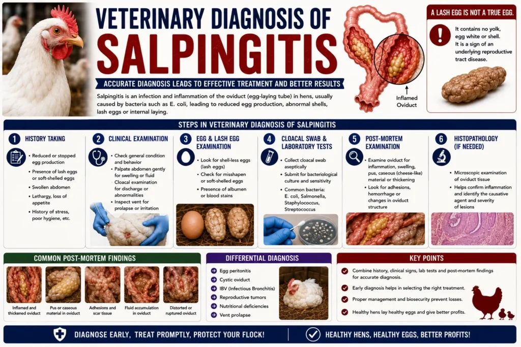
Step 1: History and Physical Examination
The vet starts by asking about laying patterns, diet, coop conditions, and recent illnesses. Then comes a hands-on exam, including gentle palpation of the abdomen to feel for firm masses, fluid, or an enlarged oviduct. The vet also checks the vent for discharge and examines the comb, weight, and overall body condition.
Step 2: Diagnostic Imaging
Radiographs (X-rays) show whether there is a mass in the abdomen and can reveal displaced organs or shell fragments from a retained egg. Ultrasound gives a clearer picture of soft tissue, helping the vet see fluid pockets or the texture of an oviduct mass. Contrast studies are sometimes used when the diagnosis remains unclear after standard imaging.
Step 3: Laboratory Testing and Bloodwork
Blood tests check white blood cell counts, since a high count usually points to infection or inflammation. A complete blood count combined with plasma biochemistry can also reveal dehydration, liver stress, or calcium imbalance. If discharge is present, a swab can be cultured to identify the exact bacteria involved, which guides antibiotic selection.
Step 4: Fine-Needle Aspiration or Coelomic Fluid Analysis
If fluid has built up in the abdominal cavity, the vet may draw a small sample with a needle. Looking at this fluid under a microscope helps separate salpingitis from egg yolk peritonitis, since the two can look identical from the outside.
Step 4: Exploratory Surgery (Laparotomy)
In borderline cases, or when treatment has not worked, a vet may recommend a small surgical incision to directly view the oviduct and ovary. This also allows a biopsy to be taken for lab confirmation.
Necropsy Findings in Fatal Cases
When a hen dies before a diagnosis is confirmed, necropsy remains one of the most definitive ways to identify salpingitis. Findings typically include a distended, thickened oviduct filled with caseous, layered material, sometimes described as looking like the inside of a hard-boiled egg cut in half. This kind of postmortem confirmation, discussed in poultry disease literature from university extension programs, helps flock owners understand what happened and adjust management for the remaining birds.
Diagnostic Methods Comparison Table
| Method | What It Shows | Invasiveness | Typical Use Case |
| Physical exam & palpation | General mass, tenderness | None | First step, every case |
| Radiograph (X-ray) | Mass location, shell fragments | Low | Suspected obstruction |
| Ultrasound | Soft tissue detail, fluid | Low | Distinguishing fluid vs. mass |
| Bloodwork/CBC | Infection markers, organ function | Low | Confirms systemic illness |
| Fluid aspiration | Fluid type/cell content | Moderate | Differentiating from peritonitis |
| Exploratory surgery | Direct visual confirmation | High | Unclear or non-responsive cases |
| Necropsy | Definitive postmortem confirmation | N/A (after death) | Sudden death cases |
Expert Tip: Bring a written timeline of symptoms and any changes in egg quality to your appointment. This speeds up the veterinary diagnosis of salpingitis because the vet can correlate physical findings with what you have already observed at home.
Differentiating Salpingitis from Similar Conditions
Because backyard keepers often self-diagnose based on symptoms alone, it helps to see how salpingitis compares with two commonly confused conditions.
| Feature | Salpingitis | Egg Yolk Peritonitis | Egg Binding |
| Location of problem | Oviduct wall | Abdominal cavity (yolk leaked) | Egg stuck in oviduct or cloaca |
| Cause | Bacterial/viral infection | Yolk released into cavity, not captured by oviduct | Weak muscle tone, large egg, calcium deficiency |
| Typical onset | Gradual | Can be gradual or sudden | Sudden |
| Fluid on ultrasound | Sometimes thick mass, less free fluid | Often free fluid present | Usually none |
| Treatment approach | Antibiotics, supportive care, sometimes surgery | Antibiotics, drainage, supportive care | Manual assistance, calcium, warmth, lubrication |
Treatment Options After Diagnosis
Treatment depends heavily on how advanced the case is at the time of diagnosis.
Medical Management
For early or mild cases, a poultry veterinarian may prescribe a course of antibiotics chosen based on culture results, along with anti-inflammatory medication and fluid therapy. Supportive care such as a warm, quiet environment and easy access to food and water plays a real role in recovery.
Surgical Intervention
Chronic or severe cases sometimes require surgical removal of the affected oviduct, a procedure called a salpingohysterectomy. This stops the hen from laying permanently but can save her life when the oviduct is too damaged to function. Surgery carries real risk in birds and should only be performed by a vet experienced with poultry.
Treatment Comparison Table
| Approach | Pros | Cons |
| Antibiotics + supportive care | Non-invasive, lower cost, no anesthesia risk | Only works in early/mild cases |
| Anti-inflammatory therapy | Reduces discomfort and swelling | Does not clear infection alone |
| Surgical removal (salpingohysterectomy) | Can resolve chronic, non-responsive cases | Higher cost, anesthesia risk, ends egg laying |
| Supportive/palliative care only | Keeps hen comfortable | Does not treat underlying infection |
Prognosis and Recovery
Hens caught in the early stage of infection, before a large mass forms, generally respond well to antibiotics and have a fair chance of returning to lay. Once caseous material has built up significantly, the prognosis becomes guarded, since this material rarely dissolves on its own and often requires surgery. Chronic cases that go untreated frequently lead to secondary complications, including peritonitis, and can be fatal.
Prevention Strategies
- Keep nest boxes clean and change bedding regularly to reduce bacterial exposure.
- Provide a balanced layer diet with adequate calcium to support strong reproductive health.
- Avoid overcrowding, which reduces stress and ammonia buildup in the coop.
- Watch body condition, since overweight hens face higher reproductive strain.
- Isolate and monitor any hen recovering from egg binding, since it raises salpingitis risk.
- Schedule a wellness check with a poultry veterinarian for hens over two years old, especially high-production breeds.
- Reduce artificial lighting extensions that push hens to lay beyond their natural rate.
Did You Know? Commercial hybrid layer breeds bred for maximum egg output are diagnosed with salpingitis more often than heritage or dual-purpose breeds, largely because of how hard their reproductive tract works over a shorter lifespan.
Common Mistakes Backyard Keepers Make
- Assuming a swollen belly is always egg binding and skipping veterinary diagnosis of salpingitis entirely
- Waiting weeks to see if symptoms resolve on their own
- Giving random antibiotics without a proper diagnosis or culture
- Ignoring subtle signs like slightly reduced appetite or one soft-shelled egg
- Not separating a sick hen from the rest of the flock during evaluation
When to Call a Poultry Veterinarian
When to Call a Vet: Contact a poultry veterinarian if a hen stops laying for more than a week without explanation, develops a swollen or firm abdomen, walks with a wide “penguin” stance, shows labored breathing, or passes foul-smelling discharge. These signs warrant a same-week appointment, not a wait-and-see approach.
Finding an experienced poultry veterinarian near you before an emergency happens makes the process faster when time matters most. Many general small-animal vets have limited experience with backyard poultry, so it helps to confirm avian or poultry experience ahead of time.
FAQ: Veterinary Diagnosis of Salpingitis
How is salpingitis diagnosed in chickens? Veterinary diagnosis of salpingitis usually starts with a physical exam and history, followed by imaging such as radiographs or ultrasound, bloodwork, and sometimes fluid aspiration or exploratory surgery to confirm the oviduct is affected.
Can salpingitis be diagnosed without a vet? Home observation can raise suspicion, but a confirmed veterinary diagnosis of salpingitis requires imaging or lab testing, since the symptoms closely resemble egg binding and egg yolk peritonitis.
Is salpingitis in hens contagious to other chickens? Salpingitis itself is not typically contagious between birds, but the underlying bacterial or viral infection causing it can sometimes spread within a flock, so isolating a sick hen is a sensible precaution.
What does a vet feel for when checking for salpingitis? A vet palpates the lower abdomen for firmness, swelling, or a mass and checks for pain response, while also assessing body condition and vent discharge.
Can a hen survive salpingitis? Many hens survive with early treatment, especially with antibiotics and supportive care. Advanced or chronic cases have a lower survival rate without surgery.
How much does a veterinary diagnosis of salpingitis typically involve in cost? Costs vary by clinic and region, but a full workup including exam, imaging, and bloodwork is often more affordable than treating a case that has progressed to needing surgery.
Does salpingitis always require surgery? No. Many early cases respond to antibiotics and supportive care. Surgery is generally reserved for chronic or non-responsive cases.