Necropsy Findings in Salpingitis: What Every Poultry Keeper and Diagnostician Should Know

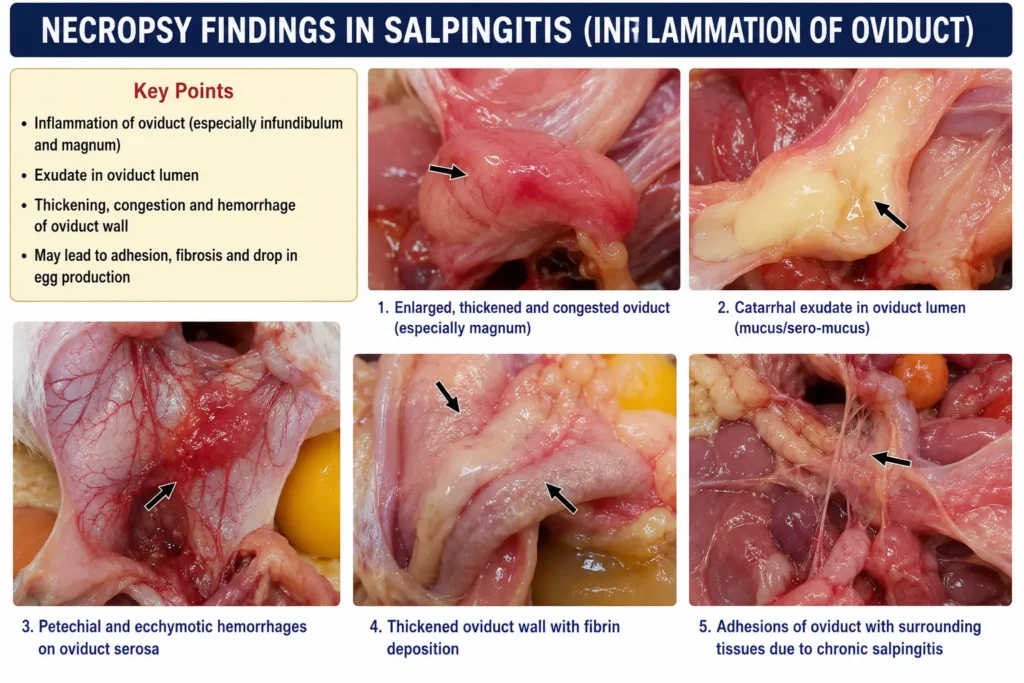
A hen that stops laying, sits puffed up in the corner, and carries a swollen belly is often hiding a problem you can only confirm after death. The necropsy findings in salpingitis are usually unmistakable once the abdomen is opened: a thickened, distorted oviduct packed with cheese-like exudate. This single lesion explains more sudden layer flock deaths than almost any other reproductive disease. This guide walks through what a postmortem chicken exam reveals, why the disease develops, and how it differs from look-alike conditions.
Table of Contents
What Is Salpingitis and Why Necropsy Matters
Salpingitis is inflammation of the oviduct, the tube that carries a developing egg from the ovary to the vent. It ranks among the top causes of death in laying hens, especially in flocks past peak production. Because the outward signs — reduced appetite, a hunched posture, occasional straining — overlap with several other illnesses, a visual diagnosis on a live bird is unreliable.
The necropsy findings in salpingitis give a far more definite answer than symptoms alone. Opening the abdominal cavity lets you see the oviduct directly, check its size and contents, and compare it against the ovary and surrounding organs. This is why veterinarians and flock managers treat postmortem exam as the gold standard for confirming the disease rather than guessing from clinical signs.
Read More: https://lashegg.com/veterinary-diagnosis-of-salpingitis/
Quick Answer
Necropsy findings in salpingitis typically include an enlarged, thickened oviduct filled with yellow-white caseous (cheese-like) material, sometimes layered like an onion, along with possible fluid in the abdomen, adhesions, and an atrophied or regressed ovary.
How Salpingitis Develops in Laying Hens
The oviduct is a long, coiled organ divided into five regions: the infundibulum, magnum, isthmus, uterus (shell gland), and vagina. Each segment adds a layer to the forming egg — albumen, shell membranes, and finally the shell. Bacteria most commonly enter through the vent (ascending infection) or spread from the bloodstream during a systemic illness.
Once bacteria establish themselves in the oviduct lining, the immune response floods the area with white blood cells. These cells, combined with mucus, cellular debris, and bacteria, dry into the thick, cheesy material pathologists call a caseous cast. This material cannot be reabsorbed or passed normally, so it accumulates and distends the organ.
Common Predisposing Factors
- Vent trauma, prolapse, or cannibalism around the cloaca
- Concurrent respiratory disease such as infectious bronchitis
- Reproductive stress from early or irregular onset of lay
- Poor litter or nest box hygiene
- Internal parasites or immune suppression from other infections
- Age — hens past 40–50 weeks show rising rates
Necropsy Findings in Salpingitis: The Core Gross Lesions
When a bird suspected of reproductive disease is opened for a postmortem chicken exam, the diagnostician works through the abdominal cavity in a set order: body condition, coelomic fluid, ovary, oviduct, and then the rest of the internal organs. The following lesions are the hallmark of salpingitis.
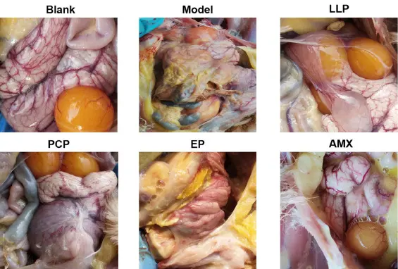
1. Distended, Thickened Oviduct
The oviduct wall loses its normal thin, glistening appearance. Instead it becomes rigid, swollen, and sometimes several times its usual diameter. In severe cases the organ can weigh many times more than a healthy oviduct.
2. Caseous Exudate
This is the single most recognizable feature among necropsy findings in salpingitis. The exudate is firm, yellow to cream colored, and often forms concentric rings when sliced, resembling a cross-section of an onion or a hard-boiled egg white gone wrong. In chronic cases it can form a solid, rubbery plug that fills the entire lumen.
3. Ovarian Changes
The ovary may appear normal early in disease, but with prolonged infection it usually regresses or atrophies because the reproductive axis shuts down. Some birds show ruptured follicles or hemorrhagic ovarian tissue if the infection has spread there directly.
4. Peritonitis and Adhesions
Because the oviduct sits within the body cavity rather than a closed tube, infection frequently spills into the peritoneum. This produces fibrinous strands, cloudy fluid, and adhesions binding the intestines, liver, and oviduct together. Pathologists call this combined picture salpingo-peritonitis.
5. Egg Yolk Peritonitis (Secondary Finding)
If yolk material escapes the reproductive tract and enters the abdomen, it triggers a strong inflammatory reaction. You may see yellow, greasy fluid coating the organs, along with a foul odor in advanced cases.
6. Enlarged, Doughy Abdomen
Before the cavity is even opened, the abdomen often feels firm, heavy, or doughy on palpation. This external clue frequently predicts what the necropsy will confirm inside.
Expert Tip: Always weigh and measure the oviduct mass when documenting necropsy findings in salpingitis. Tracking size over multiple birds in a flock helps distinguish an isolated case from an emerging outbreak.
Step-by-Step Postmortem Chicken Examination for Suspected Salpingitis
- Record history and body condition. Note age, production status, recent symptoms, and body weight before cutting.
- Open the abdominal cavity. Make a midline incision and reflect the skin and body wall to expose the coelomic organs.
- Assess fluid and odor first. Note the color, volume, and smell of any free fluid before it is disturbed.
- Locate the ovary and oviduct. Follow the reproductive tract from the ovary through to the vent, checking each segment.
- Cut open the oviduct lengthwise. Slice through the caseous material to check its layering, color, and consistency.
- Examine the liver, spleen, and air sacs. Systemic infections often leave matching lesions in these organs.
- Collect samples for bacteriology or histopathology. Submit swabs or tissue to a diagnostic lab to identify the causative organism.
- Photograph and document everything. A clear photo record supports flock-level decisions and veterinary consultation.
Did You Know?
Salpingitis is one of the most frequently detected lesions in end-of-lay hens submitted for necropsy, appearing alongside bursitis and peritonitis as a leading non-infectious and infectious cause of mortality in commercial flocks.
Table: Gross Necropsy Findings by Disease Stage
| Stage | Oviduct Appearance | Exudate | Ovary | Abdomen |
| Early / Acute | Mildly swollen, reddened lining | Thin, mucoid to slightly cloudy | Usually still active | Minimal fluid |
| Subacute | Thickened wall, partial obstruction | Soft, cream colored, patchy | Beginning regression | Mild fibrin strands |
| Chronic | Grossly distended, rigid | Firm, layered caseous cast | Atrophied or regressed | Adhesions, possible peritonitis |
| Complicated (yolk peritonitis) | Distorted, may rupture | Cast plus free yolk material | Ruptured follicles possible | Greasy yellow fluid, foul odor |
Common Bacterial Causes Behind the Lesions
Necropsy alone shows the pattern of damage, but a lab culture is needed to identify the organism responsible. The most frequently isolated bacteria include:
- Escherichia coli (E. coli) — the leading cause in commercial layers, often entering through the vent or bloodstream
- Gallibacterium anatis — associated with hemorrhagic oophoritis and peritonitis in addition to salpingitis
- Mycoplasma gallisepticum — a contributing respiratory pathogen that can predispose the reproductive tract to secondary infection
- Pasteurella multocida — linked to fowl cholera, occasionally producing reproductive lesions
- Mixed or unidentified flora — a notable share of cases show no single dominant organism on culture
Necropsy Findings in Salpingitis vs. Similar Conditions
Several other reproductive problems can mimic salpingitis on casual inspection. A careful postmortem chicken exam separates them.
| Condition | Key Distinguishing Feature | Oviduct Contents |
| Salpingitis | Caseous, layered exudate; oviduct wall thickened | Yellow-white solid cast |
| Egg-bound oviduct (impaction) | Intact, formed egg lodged in tract | Whole or partially shelled egg |
| Egg yolk peritonitis | Free yolk in abdomen, often no oviduct blockage | Minimal oviduct material; yolk on organs |
| Oviductal cancer/tumor | Firm mass with irregular texture, often unilateral | Tumor tissue, not caseous material |
| Internal laying | Multiple small yolk masses scattered in cavity | Scattered yolk, little caseous debris |
Warning
Do not assume every enlarged oviduct is salpingitis. Tumors, cystic hyperplasia, and impacted eggs can look similar on first glance. Histopathology is the only reliable way to rule out neoplasia when a mass looks unusual.
Common Mistakes When Interpreting Necropsy Findings in Salpingitis
- Skipping the ovary. Focusing only on the oviduct misses important clues about disease duration and hormonal status.
- Ignoring free abdominal fluid. Fluid color and smell often hint at whether yolk peritonitis is also present.
- Assuming one bird represents the whole flock. Necropsy several birds, not just one, before drawing flock-wide conclusions.
- Not submitting samples for culture. Gross lesions point toward salpingitis, but only bacteriology or histopathology confirms the specific cause.
- Confusing chronic scarring with active infection. Old, healed adhesions can be mistaken for a current outbreak if history is not reviewed.
When to Call a Vet
Contact a poultry veterinarian or diagnostic lab if you see repeated deaths with similar abdominal swelling, a sudden drop in flock-wide egg production, or if a home necropsy reveals oviduct changes you cannot confidently identify. A veterinarian can run bacterial culture, histopathology, and antimicrobial sensitivity testing that a backyard exam cannot replicate, and can advise on whether treatment, culling, or biosecurity changes are the right next step for the flock.
Prevention and Flock Management Implications
Necropsy findings in salpingitis are useful for more than confirming a single bird’s cause of death. Patterns across a flock guide practical management decisions:
- Reduce vent trauma risk by managing group size, lighting, and any pecking behavior.
- Support gut and reproductive health with balanced nutrition suited to the bird’s production stage.
- Control concurrent respiratory disease, since infections like infectious bronchitis raise salpingitis risk.
- Keep nest boxes and litter clean to lower environmental bacterial load.
- Cull hens with repeated laying problems rather than treating chronic cases indefinitely, since well-established lesions rarely respond to medication.
- Monitor egg production trends closely, since a flock-wide dip can be an early warning sign before mortality rises.
Antibiotics may help in the earliest stages of infection, but once the caseous cast has fully formed, medication cannot dissolve or remove it. This is why prevention and early detection through regular flock monitoring matter more than treatment once lesions are advanced.
Frequently Asked Questions
What are the main necropsy findings in salpingitis? The main necropsy findings in salpingitis are a thickened, enlarged oviduct containing firm, yellow-white caseous exudate, often with an atrophied ovary and, in advanced cases, peritonitis or adhesions in the abdomen.
Can salpingitis be diagnosed without a necropsy? Clinical signs like reduced laying, a swollen abdomen, or lethargy can raise suspicion, but a confirmed diagnosis usually requires necropsy findings in salpingitis to be directly observed, since the symptoms overlap with other reproductive diseases.
What does the caseous material in salpingitis look like? It is typically firm, cream to yellow in color, and may show concentric layers when sliced, similar in texture to a hard cheese or a cooked egg white.
Is salpingitis contagious between chickens? Salpingitis itself is not directly contagious, but the bacteria that trigger it, such as E. coli, can spread through a flock via shared environment, water, or respiratory infection, increasing the number of birds affected.
Can a hen recover from salpingitis? Early, mild cases sometimes respond to antibiotics and supportive care, but birds with an established caseous cast rarely recover fully and are usually managed by culling to prevent suffering and reduce flock risk.
What is the difference between salpingitis and egg yolk peritonitis on necropsy? Salpingitis centers on a caseous mass inside the oviduct itself, while egg yolk peritonitis shows free yolk material coating the abdominal organs, often without a solid oviduct blockage, though the two conditions can occur together.
Why is a postmortem chicken exam important for flock health? A postmortem chicken exam identifies the exact cause of death, which allows keepers and veterinarians to target the right prevention steps, whether that means improving hygiene, adjusting nutrition, or addressing a respiratory disease driving the outbreak.
Final Thoughts
Necropsy findings in salpingitis give a clear, physical answer where symptoms alone leave room for doubt. A thickened oviduct packed with caseous material, paired with ovarian regression and sometimes peritonitis, points reliably to this diagnosis. Keep monitoring your flock for early signs of reproductive trouble, keep records across multiple birds rather than a single case, and involve a poultry veterinarian whenever mortality patterns or lesions are unclear. Consistent postmortem review, alongside good hygiene and nutrition, remains the most practical way to protect flock health over time.